
EchoPixel’s True 3D Brings Holograms to Virtual Diagnostics
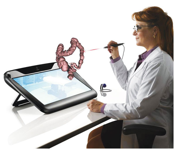
March 1, 2017 – In addition to Microsoft and their HoloLens, many other companies are creating augmented reality glasses that offer the ability to interact with holographic images. One example is EchoPixel’s FDA 510k-approved True 3D glasses. They allow radiologists to take volumetric scans from MRIs or CTs and visualize tissue and organs in a whole new way. This is paving the way for holographic-enabled virtual diagnostics (like a CT Colonography) and more efficient surgical planning.
From EchoPixelTech.com:
EchoPixel renders patient-specific anatomy in an intuitive, interactive virtual reality format, leading directly to increased clinical knowledge, faster operations, and better care. True 3D, from EchoPixel, is an advanced medical visualization software solution. It offers physicians an unprecedented opportunity to view and interact with patient tissues and organs in a truly 3D form, as if they were real physical objects.
In fact, when it comes to the clinical utility, the goals of the True 3D CT Colonography (t3D-CTC) are pretty clear:
- Improve anatomic understanding. The t3D-CTC protocol can provide a better understanding of colon lumen morphology because it integrates all three dimensions and corresponding depth cues in a single view.
- Increase polyp sensitivity. The t3D-CTC solution can provide an enhanced visualization and detection tool because the accurate depiction of depth information provided by t3D enhances the features that define a polyp (shape, size, height and edges).
- Reduce false-positive findings. With a hand-directed stylus, the reader can interact with a 2D MPR image cross section over a 3D view of a polyp or colon segment to evaluate underlying tissue density.
- Increase reader tolerance to image noise. t3D-CTC allows viewers to clearly identify 3D structures, which would have been difficult to discern from noise without accurate depiction of depth information provided by t3D.
- Reduce interpretation time. The t3D-CTC navigation strategy can eliminate anterograde and retrograde endoluminal navigation, as well as reduce reader tracking of the colon when there are redundant segments, aberrant anatomy or collapsed segments.
Today, Dr. Judy Yee at UCSF is already using the True 3D technology to help with colon cancer diagnostics.
From UCSF.edu:
At UCSF’s 3-D Imaging Lab, radiologist Judy Yee, MD, pulls up an image that looks more like a birthday party balloon animal than a patient’s colon: a vibrant, color-segmented tube, torqued and twisted in on itself.
Created from thin slices of a computed tomography (CT) scan, the image appears three-dimensional on the flat screen. It can even morph into video “fly-through” views, enhancing polyps, lesions, and other precancerous anomalies. Yee refined this revolutionary blend of advanced graphical software and scanning technology – known as CT colonography (CTC) or virtual colonoscopy – as a far less invasive and easier-to-interpret alternative to conventional colonoscopies.

Dr. Judy Yee is a professor at UCSF School of Medicine and chief of radiology at San Francisco Veterans Affairs Health Care
Yee, professor and vice chair of the Department of Radiology and Biomedical Imaging at UCSF and chief of radiology at San Francisco Veterans Affairs Health Care System, is now pushing radiology even further with holograms. Virtual holography CTC is the latest phase in her two decades of research committed to earlier, safer, life-saving detection of colorectal cancer. Though the disease is often preventable, it is the second most common cause of cancer deaths in the United States.
“When screening for breast cancer, lung cancer, prostate cancer and other malignancies, we’re typically looking for the cancer itself. By then it’s too late,” Yee says. “Here we have a specific pathology that allows us to find a lesion (known as a polyp) before it turns into cancer. If we could just get more patients to come in for screening, we could certainly have a huge impact on preventing colorectal cancer.”
Holography: Radiology’s Future?
Yee slips on a pair of 3-D black metal-rimmed glasses and points a laser stylus at the monitor still displaying her patient’s colon.
By flicking the stylus and turning her head while keeping her eyes on the monitor, Yee is suddenly “inside” the colon, moving through it, pulling it toward her, spinning it around.
Using 3-D technology, UCSF doctors can isolate and examine parts of the bowel in detail without using invasive cameras.
As she moves, a computer monitor with stereoscopic optical technology tracks her glasses, which have different polarization in each lens, prompting her brain to construct a virtual holographic object that recreates the size and shape of the human anatomy. The stylus, working in tandem with advanced graphical processing of the CTC image, allows her to “grab” the portion of the scan she wants to examine in more detail and interact with it in three-dimensional space.
“I don’t know of anyone else who is doing this,” Yee says, about UCSF’s blending of radiology with virtual holography. “You can cut away the parts of what you don’t want to see and manipulate it so that you improve what you do want to see. It’s a more engaging way to read large data sets. With the added dimension, you can see flat, more dangerous lesions better.”
The technology also has far-reaching promise for neurological, cardiac, and musculoskeletal applications, she adds.
“As the equipment evolves, it allows us to view the same disease processes in a completely different way so we can improve detection and diagnostic ability and streamline workflow,” Yee says. “This could go a long way toward helping show what radiology can bring to patient diagnosis and management for all different parts of the body.”
Find out more at echopixeltech.com
![]()

What do you think?
You are the first to add a thought